
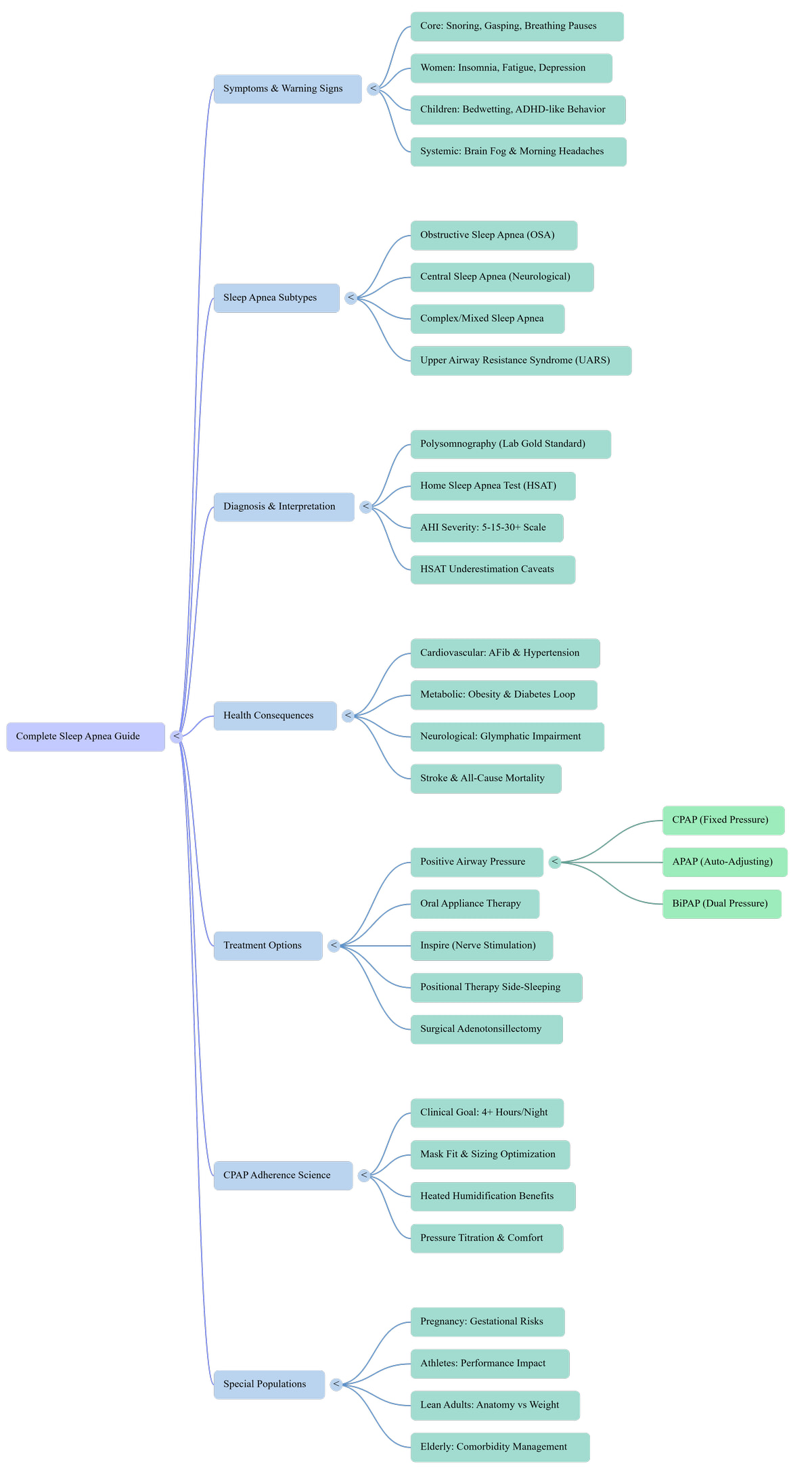
A comprehensive sleep apnea guide transforms confusion into clarity. This interactive mind map breaks down every major subtype, symptom pattern, diagnostic pathway, and treatment option into navigable layers. Unlike static images, each node reveals AI-ready explanations, evidence citations, and direct links to detailed protocols—meeting you exactly where you are in your diagnostic or treatment journey.
What does the sleep apnea guide mind map cover?
Sleep apnea guide explores seven interconnected areas: Symptoms and warning signs across genders and ages, four distinct sleep apnea subtypes including OSA and central apnea, diagnostic methods from polysomnography to home tests, cardiovascular and metabolic health consequences, treatment options from CPAP to surgical interventions, CPAP adherence science for long-term success, and special populations including pregnancy and athletes. Each area chains from recognizing symptoms through choosing evidence-based treatment.
Sleep apnea stems from repeated airway collapse or neurological signaling failure during sleep, causing oxygen drops and sleep fragmentation that drive cardiovascular disease, metabolic dysfunction, and cognitive impairment. Treatment pathways range from positive airway pressure devices to oral appliances and nerve stimulation, with adherence to therapy being the primary determinant of long-term health outcomes.
Explore Every Level of the Sleep Apnea Guide Map
This interactive sleep apnea guide invites you to explore at your own pace. Each area opens deeper—sub-topics reveal specific symptoms, diagnostic criteria, treatment mechanisms, and adherence strategies. Click any section to expand, then drill into specific techniques or read linked articles for step-by-step protocols.
Sleep apnea symptoms extend far beyond loud snoring. Core signs include witnessed breathing pauses, gasping awakenings, and non-restorative sleep despite adequate time in bed. Women often present with insomnia, fatigue, and depression rather than classic snoring, while children may show bedwetting and ADHD-like behavior. Systemic effects like brain fog and morning headaches signal oxygen deprivation during sleep.
80–90% of sleep apnea cases remain undiagnosed because symptoms present differently across genders and age groups, with many sufferers attributing fatigue to stress or aging rather than recognizing breathing disruptions (Respiratory Medicine, 2025).
Core sleep apnea symptoms center on disrupted breathing patterns. Loud, chronic snoring accompanied by witnessed pauses in breathing creates a dangerous cycle where airway collapse triggers brief awakenings to restore airflow. Gasping or choking sensations during sleep reflect the body’s emergency response to oxygen deprivation, fragmenting sleep architecture and preventing restorative deep sleep stages.
Women with sleep apnea frequently present atypically, reporting insomnia, chronic fatigue, and depression rather than loud snoring. Hormonal fluctuations during perimenopause and menopause increase vulnerability as protective progesterone declines. This atypical presentation contributes to underdiagnosis, with women waiting longer for appropriate sleep apnea diagnosis compared to men.
Pediatric sleep apnea manifests differently than adult presentations. Children may exhibit bedwetting (nocturnal enuresis), hyperactivity, inattention, and academic struggles mimicking ADHD. Enlarged adenoids or tonsils commonly cause pediatric OSA. Behavioral issues often improve dramatically after adenotonsillectomy, highlighting the importance of recognizing sleep-disordered breathing in children with behavioral or learning challenges.
Systemic sleep apnea symptoms reflect whole-body oxygen deprivation and sleep fragmentation. Morning headaches result from carbon dioxide buildup and cerebral vasodilation during apneic events. Brain fog, memory lapses, and difficulty concentrating stem from impaired glymphatic clearance—the brain’s waste removal system that operates primarily during deep sleep stages disrupted by apnea.
Sleep apnea subtypes require different treatment approaches. Obstructive sleep apnea (OSA) involves physical airway collapse affecting 83.7 million US adults. Central sleep apnea stems from neurological signaling failure where the brain fails to trigger breathing. Complex or mixed sleep apnea combines both mechanisms. Upper airway resistance syndrome (UARS) causes similar symptoms without meeting traditional AHI diagnostic thresholds, often affecting non-overweight individuals.
936 million adults globally have mild-to-severe OSA, with 425 million experiencing moderate-to-severe disease requiring intervention (The Lancet Respiratory Medicine, 2019).
Obstructive sleep apnea occurs when throat muscles relax excessively during sleep, causing the tongue, soft palate, or uvula to collapse and block the upper airway. Despite continued respiratory effort from the diaphragm and chest muscles, no airflow occurs until oxygen drops trigger a brief arousal that restores muscle tone. This cycle repeats hundreds of times nightly in severe cases, preventing restorative sleep.
Central sleep apnea results from neurological dysfunction where the brain’s respiratory control centers fail to send appropriate signals to breathing muscles. Unlike OSA, no respiratory effort occurs during apneic events. Common causes include congestive heart failure (Cheyne-Stokes breathing), stroke, opioid use, or high altitude. Treatment focuses on addressing underlying conditions and may require adaptive servo-ventilation.
Complex or treatment-emergent central sleep apnea occurs when patients with obstructive sleep apnea develop central apneas after initiating CPAP therapy. The positive airway pressure eliminates obstructions but unmasks an underlying instability in respiratory control. This subtype requires careful pressure titration and may need bilevel or adaptive servo-ventilation devices rather than standard CPAP.
Upper airway resistance syndrome causes sleep fragmentation and daytime symptoms identical to sleep apnea but without meeting traditional AHI diagnostic thresholds. Patients experience increased respiratory effort (RERAs) that trigger arousals before complete apnea occurs. UARS often affects non-overweight individuals, particularly women and young adults, and responds to similar treatments including oral appliances or low-pressure PAP therapy.
Sleep apnea diagnosis relies on polysomnography as the gold-standard in-lab test measuring brain waves, eye movements, muscle activity, heart rhythm, breathing effort, airflow, and oxygen saturation. Home sleep apnea tests (HSAT) offer convenient screening for uncomplicated cases but may underestimate severity. The apnea-hypopnea index (AHI) quantifies severity: 5-15 mild, 15-30 moderate, 30+ severe events per hour.
Polysomnography remains the gold standard for suspected OSA diagnosis; HSAT is appropriate only for uncomplicated high-probability cases, with PSG required when HSAT is negative or inconclusive (AASM Clinical Practice Guideline, 2017).
In-lab polysomnography comprehensively monitors sleep architecture, respiratory events, limb movements, cardiac rhythm, and oxygen saturation throughout the night. Trained technologists observe in real-time, allowing detection of central apneas, periodic limb movements, REM behavior disorder, and other conditions home tests miss. PSG is essential for complex cases, suspected central apnea, or when HSAT results are negative despite high clinical suspicion.
Home sleep apnea tests measure airflow, breathing effort, oxygen saturation, and heart rate using portable devices worn at home. HSAT suits uncomplicated patients with high pretest probability of moderate-to-severe OSA. However, HSAT cannot score sleep time, potentially underestimating AHI by counting recording time rather than actual sleep time, and misses central apneas, limb movements, and other sleep disorders requiring full polysomnography.
The apnea-hypopnea index quantifies sleep apnea severity by counting breathing disruptions per hour of sleep. Normal: AHI <5 events/hour. Mild: 5-15 events/hour. Moderate: 15-30 events/hour. Severe: >30 events/hour. Severity distribution shows 52% mild, 30% moderate, and 18% severe among US adults with OSA. Treatment urgency increases with severity, though even mild OSA warrants intervention if symptomatic.
Home sleep apnea tests systematically underestimate OSA severity due to methodological limitations. HSAT cannot distinguish sleep from wake, using recording time instead of sleep time for AHI calculation. Devices miss events without oxygen desaturation, fail to detect central apneas, and cannot identify sleep stage-specific or positional apnea. Negative HSAT with high clinical suspicion requires in-lab polysomnography to rule out disease.
Untreated sleep apnea triggers systemic inflammation, sympathetic nervous system overactivation, and endothelial dysfunction driving cardiovascular disease. OSA patients face five times greater hypertension risk, doubled atrial fibrillation relapse after cardioversion, and increased stroke, heart failure, and all-cause mortality. Metabolic consequences include insulin resistance and type 2 diabetes. Neurological impacts involve glymphatic impairment accelerating cognitive decline and dementia risk.
Untreated OSA patients are five times more likely to develop new-onset hypertension versus CPAP-treated patients; untreated OSA with atrial fibrillation doubles relapse risk after cardioversion (Cardiovascular Disease Risk in OSA, 2017).
Sleep apnea drives cardiovascular disease through intermittent hypoxia, intrathoracic pressure swings, and sympathetic activation. Nocturnal blood pressure surges progress to sustained hypertension. Atrial stretch and inflammation promote atrial fibrillation, with untreated OSA doubling AF relapse after cardioversion. Coronary artery disease, heart failure, and stroke risk all increase proportionally with OSA severity, making treatment essential for cardiovascular protection.
Sleep apnea and metabolic dysfunction form a vicious cycle. Intermittent hypoxia induces insulin resistance, impairs glucose tolerance, and promotes visceral fat accumulation. Leptin resistance increases appetite while sleep fragmentation reduces energy expenditure. Weight gain worsens OSA severity, which further disrupts metabolism. Breaking this cycle requires simultaneous treatment of sleep apnea and metabolic interventions including diet, exercise, and potentially diabetes medications.
Sleep apnea impairs the glymphatic system, the brain’s waste clearance mechanism operating primarily during deep sleep. Sleep fragmentation prevents sustained deep sleep stages where cerebrospinal fluid flushes metabolic waste including beta-amyloid and tau proteins. Chronic intermittent hypoxia also damages neurons and reduces neuroplasticity. These mechanisms accelerate cognitive decline, increase dementia risk, and contribute to depression and anxiety comorbidities.
Severe untreated sleep apnea significantly increases stroke risk and all-cause mortality. Mechanisms include hypertension, atrial fibrillation, endothelial dysfunction, hypercoagulability, and accelerated atherosclerosis. Nocturnal oxygen desaturation creates oxidative stress damaging blood vessels. CPAP therapy reduces cardiovascular events and mortality when used consistently ≥4 hours nightly, highlighting the life-saving potential of adherence to treatment.
Sleep apnea treatment options range from first-line positive airway pressure devices to surgical interventions. CPAP delivers fixed pressure, APAP auto-adjusts to breathing patterns, and BiPAP provides separate inspiratory and expiratory pressures for complex cases. Oral appliances advance the mandible for mild-to-moderate OSA. Inspire hypoglossal nerve stimulation prevents tongue collapse. Positional therapy addresses supine-predominant apnea. Surgical options include adenotonsillectomy and upper airway reconstruction.
CPAP therapy consistently improves cardiovascular outcomes—lowering blood pressure, reducing sleepiness scores, and decreasing serious CVD events—especially with consistent adherence ≥4 hours nightly (Is CPAP the Key to Reduce Cardiovascular Risk in OSA?, 2025).
Positive airway pressure therapy remains the gold-standard treatment for moderate-to-severe OSA. Devices deliver pressurized air through a mask to stent the airway open, preventing collapse during sleep. CPAP provides fixed pressure, APAP auto-adjusts within a range based on detected events, and BiPAP offers separate inspiratory and expiratory pressures for patients requiring higher pressures or with central apnea components.
Oral appliance therapy uses custom-fitted mandibular advancement devices to reposition the lower jaw and tongue forward, increasing upper airway dimensions and reducing collapsibility. Custom devices fitted by dentists trained in dental sleep medicine achieve better outcomes than over-the-counter options. Oral appliances suit mild-to-moderate OSA or severe OSA patients intolerant of PAP therapy, though follow-up sleep testing confirms efficacy.
Inspire hypoglossal nerve stimulation therapy uses an implanted device to prevent tongue collapse during sleep. A small generator implanted in the chest detects breathing patterns and delivers mild stimulation to the hypoglossal nerve, causing the tongue to move forward just before each inhalation. Candidates must have moderate-to-severe OSA, AHI 15-65, BMI <35, and fail CPAP therapy. Drug-induced sleep endoscopy confirms candidacy.
Positional therapy addresses supine-predominant OSA where apneas occur primarily when sleeping on the back. Gravity causes the tongue and soft palate to collapse posteriorly in supine position. Devices including specialized pillows, wearable vibratory trainers, or tennis ball techniques encourage side-sleeping. Positional therapy suits patients with >50% reduction in AHI when lateral versus supine, often combined with other treatments for comprehensive management.
Surgical adenotonsillectomy represents first-line treatment for pediatric OSA caused by enlarged tonsils and adenoids, achieving cure rates of 60-80% in children. Adult surgical options include uvulopalatopharyngoplasty (UPPP), maxillomandibular advancement, and nasal surgery. Surgery suits patients with clear anatomical obstruction, PAP intolerance, or as adjunctive therapy. Drug-induced sleep endoscopy identifies collapse sites to guide surgical planning and predict success.
CPAP adherence determines treatment success and cardiovascular protection. Clinical goals target ≥4 hours nightly use, though optimal benefit requires 6-7 hours covering full sleep periods. Mask fit and sizing optimization prevents leaks and discomfort. Heated humidification reduces nasal congestion and dryness. Pressure titration balances efficacy with comfort. Early follow-up within 30-90 days addresses barriers, with consistent users achieving 31% lower major adverse cardiovascular event risk versus inconsistent users.
Adherent CPAP use ≥4 hours nightly associates with significantly reduced major adverse cardiovascular event recurrence (HR 0.69) in OSA patients with established cardiovascular disease (JAMA IPD Meta-Analysis, 2023).
Clinical CPAP adherence goals require ≥4 hours nightly use on 70% of nights within a 30-day period for insurance compliance. However, cardiovascular and metabolic benefits scale with usage duration, with optimal outcomes at 6-7 hours covering complete sleep cycles. Even partial adherence provides some benefit versus no treatment, motivating patients struggling with full-night use to persist rather than abandon therapy.
Proper mask fit prevents air leaks causing eye irritation, noise, and ineffective therapy while minimizing skin breakdown. Mask types include nasal pillows, nasal masks, and full-face masks for mouth breathers. Sizing guides use facial measurements, but individual trial remains essential. Mask cushions require replacement every 1-3 months as silicone degrades. Side sleepers benefit from masks designed to accommodate pillow contact without dislodging.
Heated humidification adds moisture and warmth to pressurized air, preventing nasal congestion, dryness, epistaxis, and throat irritation that drive CPAP non-adherence. Heated tubing prevents rainout—condensation forming in the tube when humidified air cools. Humidity levels adjust from 1-5 based on comfort and climate. Integrated heated humidifiers significantly improve adherence compared to passive humidification or no humidification, particularly in dry climates.
Pressure titration determines optimal CPAP pressure eliminating apneas, hypopneas, and snoring while maintaining comfort. In-lab titration studies adjust pressure throughout the night across sleep stages and positions. APAP devices auto-titrate within a range, adapting to nightly variations. Ramp features gradually increase pressure as patients fall asleep. Expiratory pressure relief (EPR, Flex) reduces exhalation resistance, improving comfort and adherence for patients struggling with high pressures.
Special populations require tailored sleep apnea approaches. Pregnancy increases OSA risk through weight gain, nasal congestion, and hormonal changes, elevating gestational diabetes and preeclampsia risks. Athletes experience performance impairment from sleep fragmentation reducing VO2 max and recovery. Lean adults develop OSA from anatomical factors rather than obesity. Elderly patients face complex comorbidity management with polypharmacy interactions and cognitive impairment affecting PAP adherence.
Sleep apnea affects diverse populations beyond the stereotypical overweight middle-aged male, requiring individualized diagnostic and treatment approaches addressing unique physiological, performance, and comorbidity considerations across the lifespan.
Pregnancy increases sleep apnea risk through weight gain, upper airway edema from hormonal changes, and nasal congestion. Untreated OSA during pregnancy elevates risks of gestational diabetes, preeclampsia, gestational hypertension, and fetal growth restriction. CPAP therapy improves maternal and fetal outcomes. Screening high-risk pregnant patients with symptoms including loud snoring, witnessed apneas, or excessive daytime sleepiness enables early intervention protecting both mother and baby.
Sleep apnea impairs athletic performance through sleep fragmentation reducing VO2 max, reaction time, and recovery capacity. Intermittent hypoxia decreases oxygen-carrying capacity and mitochondrial efficiency. Athletes with OSA report decreased endurance, slower sprint times, and impaired skill execution. Treatment with CPAP or oral appliances restores sleep quality and performance metrics. Screening athletes with loud snoring, daytime fatigue, or performance plateaus identifies treatable causes of underperformance.
Lean adults develop sleep apnea from anatomical factors rather than obesity. Retrognathia (recessed chin), narrow maxilla, enlarged tongue base, or elongated soft palate narrow the airway despite normal BMI. UARS frequently affects this population. These patients often face diagnostic delays due to weight bias assuming thin individuals cannot have OSA. Treatment focuses on anatomical solutions including oral appliances, positional therapy, or surgical correction rather than weight loss.
Elderly patients with sleep apnea face complex comorbidity management including cardiovascular disease, diabetes, cognitive impairment, and polypharmacy. Age-related changes including reduced muscle tone and altered respiratory control increase OSA prevalence. Cognitive impairment affects PAP adherence requiring caregiver support. Drug interactions between sedatives, opioids, and sleep medications worsen apnea. Treatment balances cardiovascular protection with quality of life, often requiring simplified PAP interfaces and comprehensive medication review.
Going deeper? The full complete sleep apnea guide covers every protocol with step-by-step instructions.
Sleep apnea → Airway collapse or neurological signaling failure during sleep
Intermittent hypoxia → Cardiovascular disease, metabolic dysfunction, cognitive decline
CPAP adherence ≥4h/night → 31% lower cardiovascular event risk
Transform your sleep apnea diagnosis into actionable treatment success
This interactive sleep apnea guide mapped the complete landscape from symptoms through treatment adherence. The full guide provides step-by-step protocols for interpreting your sleep study, choosing between CPAP, APAP, and oral appliances, optimizing mask fit and pressure settings, and troubleshooting adherence barriers. Evidence-based strategies help you achieve the ≥4 hours nightly use that reduces cardiovascular risk by 31%. Read the complete sleep apnea diagnostic and treatment guide.
Read the Full Guide →Sources & References
This content is for informational purposes only and does not constitute medical advice. Always consult a qualified healthcare professional for personal health decisions. Content reviewed by the ZenSleepZone Research Team.